
Mouse Secondary Antibodies

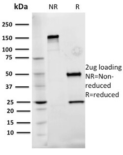
Biotium Fibronectin(Fn-3), CF488A conjugate, 0.1mg/mL
Fibronectins are disulfide-linked, dimeric glycoproteins of ~440 kDa. They possess at least four binding sites for collagen, glycosaminoglycans, transglutaminase, and a cell surface receptor. Epitope of this MAb is located in the 2nd-3rd type-III repeats of fibronectin. Fibronectins are extracellular matrix glycoproteins that are essential for embryonic development. Fibronectins are also involved in cell adhesion, tissue organization, and wound healing. Fibronectins are present in basement membranes, interstitial connective tissue matrix, and blood. Cellular fibronectin is widely distributed in the stroma of many malignant tumors. This MAb reacts with human cellular fibronectin, but not plasma fibronectin.Primary antibodies are available purified, or with a selection of fluorescent CF Dyes and other labels. CF Dyes offer exceptional brightness and photostability. Note: Conjugates of blue fluorescent dyes like CF405S and CF405M are not recommended for detecting low ab


Biotium Chromogranin A(CHGA/777), CF740 conjugate, 0.1mg/mL
Chromogranin A is present in neuroendocrine cells throughout the body, including the neuroendocrine cells of the large and small intestine, adrenal medulla and pancreatic islets. It is an excellent marker for carcinoid tumors, pheochromocytomas, paragangliomas, and other neuroendocrine tumors. Co-expression of chromogranin A and neuron specific enolase (NSE) is common in neuroendocrine neoplasms. Reportedly, co-expression of certain keratins and chromogranin indicates neuroendocrine lineage. The presence of strong anti-chromogranin staining and absence of anti-keratin staining should raise the possibility of paraganglioma. The co-expression of chromogranin and NSE is typical of neuroendocrine neoplasms. Most pituitary adenomas and prolactinomas readily express chromogranin.Primary antibodies are available purified, or with a selection of fluorescent CF Dyes and other labels. CF Dyes offer exceptional brightness and photostability. Note: Conjugates of blue fluorescent dyes
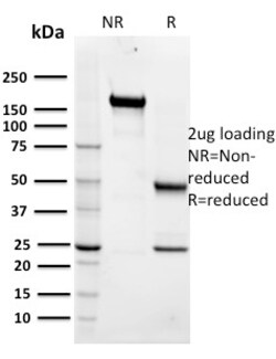
Biotium Maltose Binding Protein / MBP-probe(R29.6), CF568 conjugate, 0.1mg/mL
Plasmid vectors for the expression of coding regions of eukaryotic genes in bacterial, insect and mammalian hosts are in common usage; such expression vectors frequently encode hybrid fusion proteins consisting in part of prokaryotic and in part, eukaryotic specified proteins. One such system utilizes maltose binding protein (MBP), the 370 amino acid product of the E. coli mal E gene. Plasmid vectors have been constructed utilizing the MBP domain that allow the synthesis of high levels of MBP-fusion proteins that can be Purified in a one step procedure by affinity chromatography crosslinked amylose resin. Once bound to amylose, the MBP protein can then be separated from the target protein by cleavage by coagulation factor Xa at a specific four residue site. Alternatively, the intact fusion protein can be specifically eluted from the resin by the addition of excess free maltose. Subsequent to elution, MBP fusion protein can be visualized either by western blot analysis or immu
Biotium GFAP (Astrocyte & Neural Stem Cell Marker) (ASTRO/1974R), CF488A conjugate, 0.1mg/mL
This MAb recognizes a protein of ~50 kDa which is identified as Glial Fibrillary Acidic Protein (GFAP). It shows no cross-reaction with other intermediate filament proteins. GFAP is specifically found in astroglia. GFAP is a very popular marker for localizing benign astrocyte and neoplastic cells of glial origin in the central nervous system. Antibody to GFAP is useful in differentiating primary gliomas from metastatic lesions in the brain and for documenting astrocytic differentiation in tumors outside the CNS.Primary antibodies are available purified, or with a selection of fluorescent CF Dyes and other labels. CF Dyes offer exceptional brightness and photostability. Note: Conjugates of blue fluorescent dyes like CF405S and CF405M are not recommended for detecting low abundance targets, because blue dyes have lower fluorescence and can give higher non-specific background than other dye colors.
Biotium Ki-67 (Proliferating Cell Marker)(MKI67/2466), CF568 conjugate, 0.1mg/mL
Ki-67 antigen is a nuclear, non-histone protein that is present in all stages of the cell cycle except G0. This characteristic makes Ki-67 an excellent marker for proliferating cells and is commonly used as one of the prognostic factors in cancer studies. A correlation has been demonstrated between Ki-67 index and the histo-pathological grade of neoplasms. Assessment of Ki-67 expression in renal and ureter tumors shows a correlation between tumor proliferation and disease progression, thus making it possible to differentiate high-risk patients. Ki-67 expression may also prove to be important for distinguishing between malignant and benign peripheral nerve sheath tumors. Ki-67 labeling index has been shown to be a prognostic marker in a number of neoplasms including grade II astrocytoma, oligodendroglioma, colon carcinoma, and breast carcinoma. In general, Ki-67 is a good marker of proliferating cell populations.Primary antibodies are available purified, or with a selection of
Biotium Cytokeratin 15 (KRT15) (Basal Cell Marker) (LHK15), CF568 conjugate, 0.1mg/mL
Keratins are a family of intermediate filament proteins that assemble into filaments through forming heterodimers of one type I keratin (keratins 9 to 23) and one type II keratin (keratins 1 to 8). Keratins demonstrate tissue and differentiation specific expression profiles. Keratin 15 is a type I keratin which is expressed only in basal keratinocytes in stratified epithelia and does not appear to have a natural type II expression partner. Keratin 15 is down regulated in activated keratinocytes. Cytokeratin 15 is a specific marker of stem cells of the hair-follicle bulge and may be a useful marker for diagnosis between basal cell carcinoma (BCC) and trichoepithelioma. Trichoblastoma are benign neoplasms of follicular differentiation frequently found in nevus sebaceous. Many morphologic features are shared with nodular basal cell carcinoma, sometimes rendering a diagnosis difficult. Trichoblastoma and BCC show variable expression of Cytokeratin 15 and Cytokeratin 19, and absen
Biotium p21 / WAF1(CIP1/823), CF740 conjugate, 0.1mg/mL
This MAb recognizes a 21 kDa protein, identified as the p21WAF1 tumor suppressor protein. This MAb is highly specific to p21 and shows no cross-reaction with other closely related mitotic inhibitors. p21WAF1 is a specific inhibitor of cdk s and a tumor suppressor involved in the pathogenesis of a variety of malignancies. The expression of this gene acts as an inhibitor of the cell cycle during G1 phase and is tightly controlled by the tumor suppressor protein p53. Its expression is induced by the wild type, but not mutant, p53 suppressor protein. Normal cells generally display a rather intense nuclear p21 expression. Loss of p21 expression has been reported in many carcinomas (gastric carcinoma, non-small cell lung carcinoma, thyroid carcinoma).Primary antibodies are available purified, or with a selection of fluorescent CF Dyes and other labels. CF Dyes offer exceptional brightness and photostability. Note: Conjugates of blue fluorescent dyes like CF405S and CF405M are
Biotium DOG-1 / TMEM16A (Gastrointestinal Stromal Tumor Marker) (DG1/1485), CF568 conjugate, 0.1mg/mL
Expression of DOG-1 protein is elevated in the gastrointestinal stromal tumors (GIST s), c-kit signaling-driven mesenchymal tumors of the GI tract. DOG-1 is rarely expressed in other soft tissue tumors, which, due to appearance, may be difficult to diagnose. Immunoreactivity for DOG-1 has been reported in 97.8 percent of scorable GIST s, including all c-kit negative GIST s. Overexpression of DOG-1 has been suggested to aid in the identification of GISTs, including Platelet-Derived Growth Factor Receptor Alpha mutants that fail to express c-kit antigen. The overall sensitivity of DOG1 and c-kit in GIST s is nearly identical: 94.4% vs. 94.7%.Primary antibodies are available purified, or with a selection of fluorescent CF Dyes and other labels. CF Dyes offer exceptional brightness and photostability. Note: Conjugates of blue fluorescent dyes like CF405S and CF405M are not recommended for detecting low abundance targets, because blue dyes have lower fluorescence and can give high
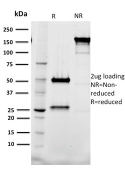
Biotium Tal1(BTL73), CF740 conjugate, 0.1mg/mL
Activation of TAL1 characterizes up to 60% of cases of human T cell acute lymphoblastic leukemia, making it the most frequent gain-of-function mutation observed in this disorder. TAL1 (also designated SCL) is a serine phosphoprotein and basic helix-loop-helix transcription factor known to regulate embryonic hematopoiesis. This transcription factor binds as a heterodimer with E2A and HEB/HTF4 to a nucleotide sequence motif termed the E-box. In addition, leukemogenesis is accelerated dramatically by transgenic co-expression of TAL1 and the catalytic subunit of casein kinase IIHLH transcription factors. Primary antibodies are available purified, or with a selection of fluorescent CF Dyes and other labels. CF Dyes offer exceptional brightness and photostability. Note: Conjugates of blue fluorescent dyes like CF405S and CF405M are not recommended for detecting low abundance targets, because blue dyes have lower fluorescence and can give higher non-specific background than oth
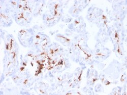
Biotium Factor XIIIa (Coagulation Factor XIIIA Chain)(F13A1/3772R), CF488A conjugate, 0.1mg/mL
Recognizes a protein of 83 kDa, which is identified as Factor XIIIa. It has been identified in platelets, megakaryocytes, and fibroblast-like mesenchymal or histiocytic cells in the placenta, uterus, and prostate, monocytes and macrophages and dermal dendritic cells. Anti-factor XIIIa has been found to be useful in differentiating between dermatofibroma (almost all cases are positive), dermatofibrosarcoma protuberans (-/ ) and desmoplastic malignant melanoma (-). Anti-factor XIIIa positivity is also seen in capillary hemagioblastoma, hemangioendothelioma, hemangiopericytoma, xanthogranuloma, xanthoma, hepatocellular carcinoma, glomus tumor, and meningioma. Primary antibodies are available purified, or with a selection of fluorescent CF Dyes and other labels. CF Dyes offer exceptional brightness and photostability. Note: Conjugates of blue fluorescent dyes like CF405S and CF405M are not recommended for detecting low abundance targets, because blue dyes have lower fluoresc
Biotium Topoisomerase II alpha (Proliferation & Drug-Resistance Marker) (TOP2A/1362), 0.2mg/mL
This antibody recognizes a 170 kDa protein, which is identified as topoisomerase IIα. It shows no cross-reaction with topoisomerase IIβ or topoisomerase I. Topo IIα plays important roles in synthesis and transcription of DNA as well as chromosomal segregation during mitosis. It is reported to be a sensitive and specific marker of late S-, G2- & M-phases in transformed and developmentally regulated normal cells. Topo IIα is also implicated in drug resistance of tumor cells.Primary antibodies are available purified, or with a selection of fluorescent CF Dyes and other labels. CF Dyes offer exceptional brightness and photostability. Note: Conjugates of blue fluorescent dyes like CF405S and CF405M are not recommended for detecting low abundance targets, because blue dyes have lower fluorescence and can give higher non-specific background than other dye colors.
Biotium Smooth Muscle Actin(ACTA2/791), CF740 conjugate, 0.1mg/mL
Actin is a major component of the cytoskeleton and is present in most cell types. It is highly specific to actin from smooth muscles. This MAb does not stain cardiac or skeletal muscle; however, it does stain myofibroblasts and myoepithelial cells. This antibody could be used together with anti-muscle specific actin and myogenin in making a diagnosis of smooth muscle and skeletal muscle tumors. In most cases of rhabdomyosarcoma, this antibody yields negative results whereas anti-muscle specific actin and myogenin are positive. Leiomyosarcomas are positive only with anti-muscle specific actin and anti-smooth muscle actin and are negative with anti-myogenin.Primary antibodies are available purified, or with a selection of fluorescent CF Dyes and other labels. CF Dyes offer exceptional brightness and photostability. Note: Conjugates of blue fluorescent dyes like CF405S and CF405M are not recommended for detecting low abundance targets, because blue dyes have lower fluoresce
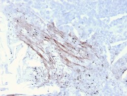
Biotium Tenascin C (Stromal Marker for Epithelial Malignancy)(TNC/3635), CF568 conjugate, 0.1mg/mL
In Western blotting, this antibody reacts with two bands of ~MW of 210 kDa and 300 kDa, identified as two isoforms of Tenascin C. Specificity of this MAb is validated by sequential immunoprecipitation with a PAb against Tenascin C. Tenascin C is a multifunctional, disulfide-linkedhexameric extracellular matrix glycoprotein expressed in association with mesenchymal epithelial interactions during development and in the neo-vasculature and stroma of undifferentiated tumors. In adults, it is restricted to certain epithelial-stromal interfaces and increases markedly in hyper-proliferative diseases and in stroma of many neoplasms, including gliomas, breast, squamous and lung carcinomas. Primary antibodies are available purified, or with a selection of fluorescent CF Dyes and other labels. CF Dyes offer exceptional brightness and photostability. Note: Conjugates of blue fluorescent dyes like CF405S and CF405M are not recommended for detecting low abundance targets, because blue
Biotium MART-1 / Melan-A(MLANA/788), CF740 conjugate, 0.1mg/mL
This antibody recognizes a protein doublet of 20-22 kDa, identified as MART-1 (Melanoma Antigen Recognized by T cells 1) or Melan-A. MART-1 is a newly identified melanocyte differentiation antigen recognized by autologous cytotoxic T lymphocytes. Seven other melanoma associated antigens recognized by autologous cytotoxic T cells include MAGE-1, MAGE-3, tyrosinase, gp100, gp75, BAGE-1, and GAGE-1. Subcellular fractionation shows that MART-1 is present in melanosomes and endoplasmic reticulum. This MAb labels melanomas and other tumors showing melanocytic differentiation. It is also a useful positive-marker for angiomyolipomas. It does not stain tumor cells of epithelial, lymphoid, glial, or mesenchymal origin.Primary antibodies are available purified, or with a selection of fluorescent CF Dyes and other labels. CF Dyes offer exceptional brightness and photostability. Note: Conjugates of blue fluorescent dyes like CF405S and CF405M are not recommended for detecting low abu
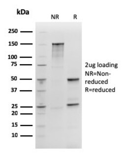
Biotium SOX2 (Embryonic Stem Cell Marker)(rSOX2/1792), Biotin conjugate, 0.1mg/mL
SOX2 is required for stem cell maintenance in the central nervous system, and it also regulates gene expression in the stomach. SOX2 is necessary for regulating multiple transcription factors that affect Oct 3/4 expression. An essential function of SOX2 is to stabilize embryonic stem cells in a pluripotent state by maintaining the requisite level of Oct 3/4 expression. Reportedly, SOX2 is associated with aggressive phenotypes of breast, head and neck, gastric, colorectal, bladder, and small cell lung cancers. However, SOX2 is expressed in a high percentage of lung squamous cell carcinomas and has been shown to be an independent favorable prognostic marker. Primary antibodies are available purified, or with a selection of fluorescent CF Dyes and other labels. CF Dyes offer exceptional brightness and photostability. Note: Conjugates of blue fluorescent dyes like CF405S and CF405M are not recommended for detecting low abundance targets, because blue dyes have lower fluoresc